
When you look at your own face in a mirror, or when a surgeon examines your face during a standard rhinoplasty consultation, what's actually being assessed is the visible surface, skin, soft tissue contour, and overall proportion as it currently appears. A cephalometric X-ray provides something genuinely different: a standardized, measurable view of the underlying bony structure itself, the actual skeletal framework beneath everything visible, along with, in some cases, key soft tissue landmarks positioned relative to that skeletal foundation. This is the blueprint beneath the finished structure, and for certain surgical planning decisions, this underlying blueprint matters as much or more than the visible surface it supports.
Why "Cephalometric" Specifically, and Not Just Any X-Ray
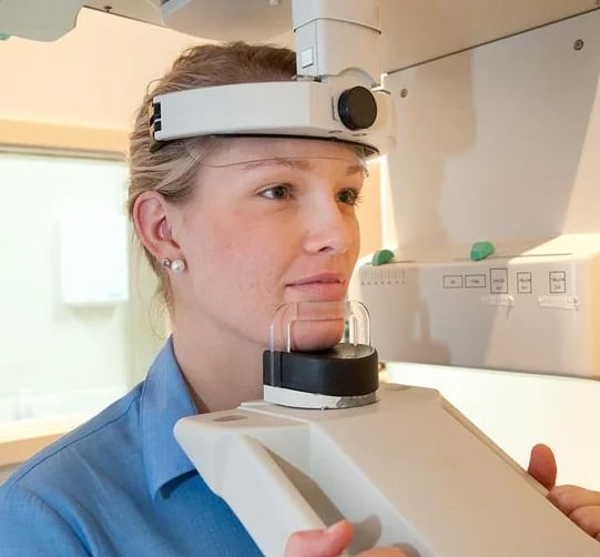
The term cephalometric refers to a specific, standardized imaging technique, distinct from a general skull X-ray, performed with the patient's head positioned in a precisely calibrated, reproducible orientation using a device called a cephalostat, which holds the head in an exact, consistent position relative to the X-ray source and film or digital sensor. This standardization matters enormously, since it allows the resulting image to be measured with genuine precision and, importantly, compared meaningfully against established population norms or against a patient's own prior imaging if planning involves tracking change over time, neither of which would be reliably possible with a less standardized imaging approach where head position varies between images.
What Specific Measurements the Blueprint Actually Reveals
Once captured, a cephalometric X-ray allows a surgeon to take specific, precise measurements of skeletal relationships that aren't reliably assessable through visual examination alone, including the relative position and projection of the upper and lower jaw to each other and to the rest of the facial skeleton, the specific angle and position of the chin relative to established facial landmarks, and the underlying bony support for the nose, relevant specifically to rhinoplasty planning in patients whose nasal profile is influenced significantly by underlying skeletal structure rather than soft tissue and cartilage alone.
These aren't measurements a trained eye can reliably estimate through visual assessment, even an experienced, skilled visual assessment, since the actual skeletal position is genuinely obscured by the overlying soft tissue, muscle, and skin that varies in thickness from patient to patient, sometimes creating a visible profile that doesn't directly, transparently reflect the underlying bony structure supporting it.
Where This Blueprint Matters Most: Combined Chin and Nose Planning
This specific imaging tool becomes particularly valuable in exactly the kind of combined chin-and-nose planning discussed in relation to overall facial proportion elsewhere in our content, since accurately assessing whether a nose appears disproportionate due to its own structure, or due to an underprojected chin creating a relative appearance of nasal prominence, genuinely benefits from precise skeletal measurement rather than visual impression alone. A cephalometric X-ray allows a surgeon to determine with real precision whether a specific patient's chin sits behind an established reference line relative to their actual skeletal structure, information that directly informs whether chin augmentation, rhinoplasty, or a combination of both represents the most appropriate surgical plan for that specific patient's actual underlying anatomy.
Where This Blueprint Matters: Complex or Revision Rhinoplasty Cases
For patients considering revision rhinoplasty, where the nose's original structure has already been surgically altered once and the relationship between remaining bone, cartilage, and soft tissue may be less straightforward to assess visually than in a primary, unoperated case, cephalometric imaging can provide valuable additional information about the underlying skeletal framework that visual examination and even standard photography don't fully capture. This additional information becomes particularly relevant when planning a revision procedure specifically needs to account for what skeletal support genuinely remains available to work with, following whatever structural changes the original surgery already made.
Where This Blueprint Isn't Necessary: Straightforward Cosmetic Cases
It's worth being direct that cephalometric imaging isn't a standard, universal component of every facial surgery consultation, and appropriately so. Many straightforward cosmetic rhinoplasty and chin procedures, particularly in patients with relatively uncomplicated anatomy and clear, well-defined aesthetic goals, are planned effectively through standard visual examination, photography, and, in some practices, digital imaging software allowing patients and surgeons to visualize potential changes, similar to the planning conversation for a facelift consultation where soft tissue assessment alone is often sufficient, without requiring this additional, more specialized imaging step. Cephalometric analysis becomes specifically valuable in the more complex cases described above, not as a routine, universal requirement for every patient considering any facial procedure.
What the Actual Imaging Appointment Involves
For patients whose surgical plan does call for this specific imaging, the appointment itself is brief and non-invasive, involving positioning in the cephalostat device for a matter of seconds while the image is captured, using a radiation dose comparable to other standard dental or medical X-ray imaging, well within established safety guidelines for this kind of diagnostic imaging. This isn't a significant additional burden added to your surgical planning process, it's a brief, specific step that provides genuinely valuable additional information for the particular subset of cases where it meaningfully changes or confirms the surgical plan.
How This Blueprint Actually Changes a Surgical Recommendation in Practice
Consider a patient presenting with a concern focused entirely on their nose, requesting rhinoplasty to reduce what they perceive as excessive nasal prominence. A cephalometric analysis in this specific case might reveal that this patient's nasal bone and cartilage structure actually falls within a fairly typical, unremarkable range, while their chin sits meaningfully behind the established reference line discussed earlier, meaning the perceived nasal prominence is substantially a relative effect created by underprojection elsewhere in the facial profile, not primarily an issue with the nose's own underlying structure. This kind of finding can meaningfully redirect a surgical conversation toward chin augmentation alone or a more conservative rhinoplasty combined with chin augmentation, rather than the more extensive nasal reduction the patient may have initially assumed they needed based on visual impression alone.
Why This Level of Precision Reflects a Broader Approach to Surgical Planning
This specific imaging tool represents one part of a broader philosophy worth understanding directly: that facial surgical planning benefits from objective, measurable information wherever it's genuinely available and relevant, rather than relying exclusively on visual assessment and aesthetic judgment, however skilled, when a more precise underlying picture would meaningfully inform the surgical plan. This same principle underlies the careful, structural approach Dr. Doshi brings to preservation rhinoplasty technique more broadly, where understanding a patient's actual underlying structure precisely, rather than working from visual impression alone, directly shapes how conservatively or extensively a given procedure needs to address that structure to achieve the intended result.

What a Consultation Involving This Imaging Should Actually Cover
For patients whose consultation includes cephalometric imaging as part of their surgical planning, a thorough process should include a clear explanation of why this specific imaging is being recommended for your particular case, what specific measurements or relationships it's expected to clarify, and how the resulting findings will directly inform your surgical plan, rather than the imaging being performed as an unexplained, generic step without this specific context.
Dr. Doshi incorporates cephalometric analysis specifically for the complex cases described throughout this piece, using this objective measurement to inform decisions around combined chin and nasal planning or revision rhinoplasty where underlying skeletal structure genuinely changes the appropriate surgical approach. This same precision-first philosophy extends to his broader approach to primary rhinoplasty planning generally.
Patients earlier in their research process can review our overview of surgical facial procedures for a broader picture of how this kind of precise, structural planning fits into the overall consultation and surgical process.