
Cheek dimples occupy a particular place in the aesthetics of the face. They are associated across many cultures with attractiveness, youthfulness, and expressiveness, and they have a striking quality in that they appear only with certain facial expressions. A dimpled smile looks different from a smile without dimples in a way that is immediately recognisable and almost universally considered appealing.
For people who were not born with dimples and want them, the relevant question is whether they can be created surgically in a way that looks natural, lasts, and carries acceptable risk. The answer to all three is yes, but the details of each matter, and most patients who search for dimpleplasty on Long Island arrive without a clear picture of what the procedure actually involves, what the result looks like at one week versus one year, and what the honest limitations are.
Dr. Hardik Doshi is a double board-certified facial plastic surgeon at Doshi Plastic Surgery in Garden City, Long Island and Manhattan. He performs dimpleplasty as part of his specialty procedures practice alongside facial plastic surgery, and has a documented gallery of results for Long Island patients. What follows is a complete guide to dimpleplasty: the anatomy, the technique, the healing timeline, the realistic result, and the questions every patient should have answered before booking.

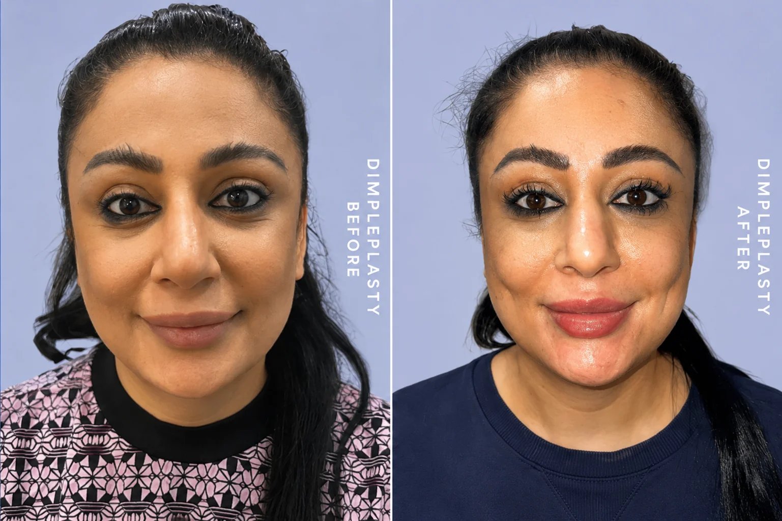
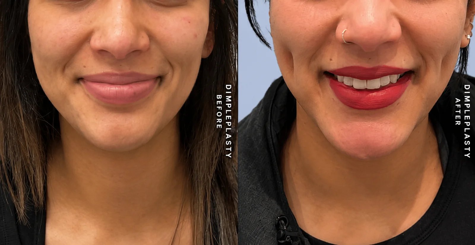
Patients who want to see documented results before their consultation can review the dimpleplasty before and after gallery at Doshi Plastic Surgery. The dimpleplasty procedure page provides a summary of what the procedure involves.
Why Some People Have Natural Dimples and Others Do Not
Understanding why natural dimples form clarifies what dimpleplasty is actually replicating. Natural cheek dimples are caused by a variation in the anatomy of the zygomaticus major muscle, the primary muscle responsible for pulling the corner of the mouth upward during smiling. In people with natural dimples, this muscle is bifid, meaning it has a split or double-headed configuration rather than a single continuous muscle belly.
When a bifid zygomaticus major contracts during smiling, the split creates a point of attachment to the overlying skin between the two muscle heads. The skin is pulled inward at this point, creating the characteristic indentation of the dimple. The dimple is visible with animation, specifically with the facial expressions that engage the zygomaticus major, and is absent or much less visible at rest in most people with natural dimples.
Dimpleplasty replicates this mechanism artificially. Instead of a bifid muscle creating an attachment to the skin, the procedure creates a controlled adhesion between the inner surface of the cheek and the dermis of the overlying skin, through which the skin is tethered and dimples when pulled by the facial musculature during animation. The biological mechanism of the result is the same as in natural dimples. The method of creating it is different.
The Anatomy of the Cheek and What the Procedure Works With
The cheek has several anatomical layers between the skin surface and the oral mucosa. From outside to inside: the skin, a layer of subcutaneous fat of variable thickness depending on the patient's facial fullness, the buccinator muscle, which forms the muscular wall of the cheek and plays a primary role in keeping food between the teeth during chewing, a thin fascial layer, and the oral mucosa lining the inside of the mouth.
Dimpleplasty creates an adhesion between the dermis (the deeper layer of the skin) and the buccinator muscle or its overlying fascia. This adhesion, when it heals, tethers the skin at the dimple point so that when the surrounding facial muscles contract during smiling or animation, the tethered skin is held in place relative to the surrounding tissue, creating the inward indentation that reads as a dimple.
The buccinator is a relatively thin, flat muscle. It is not a structure that is significantly altered by the procedure. The suture that creates the adhesion catches the superficial surface of the muscle or its fascial covering rather than passing through the muscle itself. This is one reason the procedure carries a low complication profile when performed with correct anatomical knowledge.
Cheek fullness is relevant to how the dimple appears. In patients with very full cheeks, a significant depth of subcutaneous fat separates the skin from the buccinator, which can make the dimple less defined because the tethering adhesion has more tissue to pull through before the surface indentation becomes visible. In patients with leaner facial anatomy and less subcutaneous fat in the cheek, the dimple may be more clearly defined. This is a factor in the consultation discussion, not a contraindication, but it affects realistic expectations about definition and depth.
How Dimpleplasty Is Performed
Dimpleplasty is an office procedure performed under local anaesthesia. There are no general anaesthesia requirements, no operating room booking, and no IV lines. The patient sits in a treatment chair, the cheek is cleaned with an antiseptic solution, and local anaesthetic is injected both from the outside and from inside the mouth to numb the full thickness of the cheek.
Marking the Position
Before any instrumentation, the dimple position is marked on the skin surface. This is one of the most important steps in the procedure and one where surgeon judgment and understanding of facial aesthetics matter considerably.
The marking is done with the patient smiling. The surgeon identifies the location on the cheek where a dimple would most naturally and attractively complement the patient's specific facial anatomy. This involves assessing the relationship between the cheek, the nasolabial fold, the corner of the mouth, and the overall proportions of the face. Most commonly, the dimple is marked along a line extending from the corner of the mouth to the tragus of the ear, but the precise position along this line and the lateral-to-medial placement are individualised.
Patients are encouraged to bring photographs of dimple positions they find attractive, not necessarily to replicate them exactly, but to communicate the aesthetic they are drawn to. The final position is agreed upon with the patient before any instrumentation begins.
Creating the Adhesion
Once the position is marked and the anaesthetic has taken effect, a small biopsy punch or fine instrument creates a tiny entry point through the oral mucosa from inside the mouth, at the point corresponding to the marked skin position. This approach avoids any external skin incision and leaves no visible external scar.
A suture is passed through this entry point and positioned to catch a small amount of the buccinator muscle fascia and the dermis of the overlying skin simultaneously. When this suture is tied, it draws the dermis down toward the muscle, creating the initial tethering adhesion. The suture material used is typically absorbable, meaning it does not need to be removed later. The dimple is formed by the fibrous adhesion that develops as healing occurs around the suture, not by the suture itself remaining permanently in place.
The entry point inside the mouth is either left to heal on its own or closed with a single absorbable suture. There is no external wound to care for. The patient leaves with an immediately visible dimple and mild local swelling.
Bilateral vs Unilateral Dimpleplasty
Most patients request bilateral dimpleplasty, creating a matching dimple on each cheek. The procedure is performed sequentially, with each side requiring the full marking, anaesthesia, and instrumentation process. The total procedure time for bilateral dimpleplasty is approximately 45 to 60 minutes.
Some patients request a unilateral dimple on one side only. This is a less common request but is performed for patients who have a natural dimple on one side and want symmetry, or who simply prefer the aesthetic of a single cheek dimple. There is no clinical reason to require bilateral dimpleplasty if the patient's preference is for one side.
The Healing Timeline: What to Expect at Each Stage
The healing process after dimpleplasty follows a predictable sequence, and understanding it in advance prevents the anxiety that can arise when patients see their result looking different from what they expected in the early weeks.
Days One to Seven: The Dimple Appears Constantly
In the first week after dimpleplasty, the dimple is visible all the time, both at rest and with animation. This is not the final result. The constant visibility at rest during this period reflects the initial swelling and inflammation around the adhesion site, which holds the tissue in a tethered position regardless of facial expression. The cheek is mildly swollen, and the dimple may appear deeper than it will ultimately be as the surrounding tissue settles.
The inside of the mouth may have mild soreness at the entry point. Eating is comfortable but patients are advised to stay on soft foods for the first few days to reduce mechanical stress on the healing site. Rigorous chewing, particularly of hard or sticky foods, is discouraged during the first week.
Oral hygiene is important throughout the healing period. Patients are advised to rinse the mouth after eating to keep the intraoral entry point clean. Standard mouthwash is typically introduced at 48 hours.
Weeks Two to Four: The Dimple Transitions
As the initial swelling resolves, the dimple begins its transition from a constantly visible indentation to one that appears primarily with animation. By two to three weeks, most patients find that the dimple is still visible at rest but is becoming less pronounced in repose while remaining well-defined with smiling. The cheek swelling is largely resolved, and the patient is comfortable in social settings.
This transitional phase can be slightly disconcerting for patients who expected the constant dimple of the first week to represent the permanent result. It does not. The dimple is healing toward its intended behaviour: dynamic visibility with animation, with a subtle shadow or indentation at rest.
Months One to Three: The Result Settles
By one to three months, the fibrous adhesion that gives the dimple its permanence has matured. The dimple at this stage is visible primarily with facial animation and shows a subtle but perceptible indentation at rest in most patients. The exact degree of at-rest visibility varies between individuals depending on their healing response, the depth of the initial adhesion, and their facial anatomy.
This is the stage at which before and after photography is most informative. The result at three months represents approximately where the final result will be, and patients can assess whether the position, depth, and behaviour of the dimple meets their expectations.
Long-Term: Permanence and Natural Behaviour
The fibrous adhesion created by dimpleplasty is intended to be permanent. Unlike muscle relaxing injections or fillers, which metabolise and require maintenance, the dimple is held by fibrous tissue that does not dissolve. The dimple created at surgery will behave with the face as the face ages, moving naturally with facial expression and changing proportionally with any changes in facial fullness over time.
In patients who gain significant weight after dimpleplasty, increased cheek fullness may make the dimple less visible. In patients who lose significant facial volume with age, the dimple may become more pronounced. These are natural consequences of the dimple behaving as part of the face's living anatomy.
Choosing the Right Position: The Most Important Decision in the Procedure
Of all the variables in dimpleplasty, the position of the dimple has the greatest influence on whether the result looks natural and attractive or placed. A dimple in the wrong position, even a technically perfect dimple, will not look like a natural dimple. It will look placed.
Natural dimples occur in a relatively predictable anatomical territory, typically along a line from the oral commissure to the tragus and positioned at a point that corresponds to where the zygomaticus major muscle lies beneath the skin. Dimples placed outside this zone, particularly dimples placed too far lateral toward the ear or too low toward the jaw, do not replicate the natural pattern and read as artificial.
The consultation assessment includes direct observation of the patient smiling, speaking, and making the range of facial expressions that animate the cheeks. The surgeon watches how the cheek moves with animation, identifies the point where a dimple would fall naturally given the patient's specific facial dynamics, and marks this position before the patient makes a final decision.
Patients who arrive with specific reference photographs of dimple positions they find attractive are encouraged to share them. This is genuinely useful information for the consultation. It is not that the reference dimple will be replicated precisely, but that understanding what the patient finds aesthetically attractive about a particular dimple position helps the surgeon ensure the final placement reflects the patient's taste rather than a generic anatomical average.
Combining Dimpleplasty With Other Procedures
Dimpleplasty is frequently booked as a standalone procedure, but it is also commonly combined with other facial aesthetic treatments at Doshi Plastic Surgery when patients are addressing multiple concerns.
Patients who are having buccal fat removal to slim the midface often consider adding dimpleplasty to complement the reduced cheek fullness with a defined dimple. The combination of leaner cheek anatomy and a well-placed dimple can produce a result that looks particularly defined and sculpted.
Patients considering chin augmentation or lip lift surgery occasionally add dimpleplasty to the same appointment, as the procedures do not conflict and share a recovery period. The ability to address multiple facial aesthetic concerns in a single session is a practical consideration that patients managing time and recovery preference appreciate.
For patients considering non-surgical facial enhancement alongside dimpleplasty, dermal filler treatments for cheek volumising or lip augmentation can be scheduled at the same appointment with appropriate sequencing.
Risks and Complications: The Honest Account
Dimpleplasty is a low-complexity procedure with a low serious complication rate when performed by an experienced practitioner with correct anatomical knowledge. However, as with any surgical procedure, risks exist and should be understood before booking.
Asymmetry
The most common outcome concern in dimpleplasty is asymmetry between the two dimples in bilateral cases. This can reflect a difference in depth, position, or the degree of at-rest visibility between the two sides. Minor asymmetry is common in natural dimples and is not always perceived as a problem. Significant asymmetry that is visibly uneven can be addressed through a revision procedure to adjust the less satisfactory side.
Dimple Too Deep or Too Shallow
The depth of the dimple depends on the depth of the initial adhesion and the thickness of the subcutaneous fat between the skin and the buccinator. If the dimple is too deep, it may appear exaggerated rather than natural. If the adhesion is insufficient, the dimple may be too subtle to be satisfying. The surgeon's judgment about suture placement and tension at the time of the procedure is the primary determinant of depth. Patients should review before and after photographs of the specific surgeon's results before proceeding.
Infection
The procedure creates a pathway between the inside of the mouth, which carries a normal bacterial flora, and the cheek tissue. Infection is uncommon because the mouth heals efficiently and the tissue has good vascular supply, but it is a recognised risk. Patients are typically given a brief course of oral antibiotics post-operatively to reduce infection risk, and meticulous oral hygiene during healing further minimises it.
Visible Internal Suture
Occasionally, an absorbable suture placed during dimpleplasty can become palpable or visible inside the mouth before it fully dissolves. This is a temporary phenomenon that resolves as the suture material resorbs. If a suture becomes symptomatic or bothersome before it resorbs on its own, it can be removed in the office.
The Question of Permanence
Dimpleplasty is permanent in intention. Some patients, having had the procedure, find that they wish to have the dimple reduced or removed. Early release of the adhesion in the first weeks, before full fibrosis has occurred, is possible but not always completely reversible. Once the fibrous adhesion has fully matured, reversal involves a small procedure to surgically release the adhesion, the outcome of which is unpredictable in terms of how completely the original appearance is restored.
This is the most important pre-operative consideration: dimpleplasty is a permanent procedure. The decision to have it should reflect considered, settled preference rather than a transient interest, and the patient should be genuinely comfortable with the prospect of a permanent change to their facial appearance.
Who Is the Right Patient for Dimpleplasty on Long Island
The right candidate for dimpleplasty at Doshi Plastic Surgery is a patient who:
- Has a settled, genuine desire for cheek dimples that reflects their longstanding aesthetic preference rather than a recent trend-driven interest.
- Understands and accepts that the result is intended to be permanent.
- Has realistic expectations about the depth and visibility of the dimple based on their specific facial anatomy, including their cheek fullness.
- Is in generally good health with no conditions that significantly impair healing or increase infection risk.
- Has no active oral or skin infections in or adjacent to the treatment area.
- Is prepared to follow post-operative instructions including dietary modification and oral hygiene protocols during the healing period.
There is no single ideal age or facial type for dimpleplasty. The procedure is most commonly performed in patients in their 20s and 30s, but appropriately selected patients outside this range are also good candidates. Cheek fullness affects the depth and definition of the result and is discussed explicitly at consultation.
What to Expect at the Dimpleplasty Consultation at Doshi Plastic Surgery
The consultation begins with Dr. Doshi examining the patient's facial anatomy while the patient is both at rest and during animation. He observes how the cheeks move with smiling and expression, identifies the anatomical territory where a dimple would naturally fall for that patient's specific face, and discusses the proposed position with the patient before making any marks.
Patients are shown before and after photographs of dimpleplasty results from Dr. Doshi's own practice, including cases where the patient's facial anatomy is similar to theirs. The consultation includes a direct discussion of what the result will look like at one week, at one month, and at one year, so the patient understands the healing progression before they encounter it.
Any questions about the procedure, the recovery, the permanence of the result, or the risk of asymmetry or other complications are addressed directly. Patients are encouraged to take time to think after the consultation before booking, particularly given the permanent nature of the change.
For patients who are not ready for an in-person consultation, virtual consultations are available as a first step. Scheduling is available through doshiplasticsurgery.com or by contacting the practice directly.
Why Dr. Doshi on Long Island
Dimpleplasty is one of those procedures where the line between an experienced facial surgeon and a less experienced practitioner matters more than the procedure's simplicity might suggest. The position of the dimple is an aesthetic judgment that requires genuine understanding of facial anatomy and proportion. The depth and tension of the adhesion suture is a surgical judgment that determines whether the result is natural or exaggerated. And the complication management, including the ability to recognise and address asymmetry or an infection, requires clinical experience. Dr. Doshi's background as a double board-certified facial plastic surgeon, detailed on the about Dr. Doshi page, means that every dimpleplasty at Doshi Plastic Surgery is performed by a surgeon whose entire training and practice is focused on the anatomy and aesthetics of the face.
Doshi Plastic Surgery is located in Garden City, Long Island, with a second office in Manhattan. Patients from across Nassau County, Suffolk County, Queens, and Brooklyn regularly attend the practice for procedures not readily available at other Long Island facilities. Dimpleplasty is one of these procedures, and the gallery at the practice documents results across a diverse range of patients and facial types.
What is dimpleplasty and how does it work?
Dimpleplasty is a minimally invasive surgical procedure that creates a permanent cheek dimple by forming a controlled adhesion between the inner surface of the cheek mucosa and the underlying facial muscle. A small instrument is introduced through a tiny incision inside the mouth, and a suture is placed that tethers a small amount of the buccinator muscle to the overlying dermis. When this adhesion heals, it creates an indentation in the skin surface that mimics the appearance of a natural dimple. The procedure involves no external incisions and leaves no visible scars.
Are dimpleplasty results permanent?
Dimpleplasty produces results that progress through a predictable sequence. In the first weeks after surgery, the dimple is visible both at rest and with animation. Over the following weeks to months, the dimple typically becomes visible primarily with animation, when smiling or speaking, rather than constantly at rest. In most patients, a permanent subtle indentation or shadow remains even at rest over time, while the dimple deepens and becomes more defined with facial movement. The long-term result varies by individual healing response and the depth of the initial adhesion.
Where exactly are cheek dimples placed during dimpleplasty?
Dimple placement is determined through a consultation assessment that considers the patient's facial anatomy, cheek fullness, natural smile dynamics, and aesthetic preference. The most common placement follows a line from the corner of the mouth to the tragus of the ear, with the dimple positioned at the point where it would most complement the patient's specific facial proportions. The position is marked on the skin surface while the patient is smiling to confirm that the selected location produces a natural-looking indentation with animation.
Is dimpleplasty painful?
The procedure is performed under local anaesthesia, which numbs the cheek tissue thoroughly before any instrumentation. Patients feel pressure but no pain during the procedure itself. Post-operative discomfort is typically mild, described as similar to the soreness following a dental procedure. Over-the-counter analgesics are generally sufficient for the first few days. Swelling in the treated cheek is the dominant post-operative symptom and is visible for the first one to two weeks.
How long does dimpleplasty take and what is the recovery like?
The procedure takes approximately 20 to 30 minutes per side in an office setting. Recovery involves mild to moderate swelling in the treated cheek for one to two weeks, during which the dimple appears constantly visible rather than only with animation. Most patients return to normal daily activities the day after surgery. A soft diet is recommended for the first few days. Social settings are typically comfortable again at ten to fourteen days.
Can dimpleplasty be reversed if I change my mind?
Dimpleplasty is considered a permanent procedure. Early in the post-operative period, before the adhesion has fully matured, some surgeons can release the suture and reduce the prominence of the dimple. Once the fibrous adhesion has fully formed and matured, typically after several months, reversal is more complex and may involve a small procedure to release the adhesion, with no guarantee of complete return to the original appearance. Patients should consider dimpleplasty a permanent change and approach the decision accordingly.
Who is a good candidate for dimpleplasty on Long Island?
Good candidates are patients in generally good health with realistic expectations about the nature of the result who have considered their decision carefully and understand that the procedure creates a permanent change. There is no ideal facial type or age range, though the procedure is most popular in patients in their 20s and 30s. Patients with fuller cheeks may have a less defined dimple than patients with leaner facial anatomy, and this should be discussed at consultation.
Are dimples from dimpleplasty visible all the time or only when smiling?
The appearance of dimpleplasty results changes over time. In the first weeks post-operatively, the dimple is typically visible both at rest and with animation due to the initial adhesion and healing inflammation. As healing progresses over weeks to months, most patients find their dimple becomes primarily visible with animation, particularly when smiling or speaking. A subtle indentation or shadow may remain visible at rest. The dynamic dimple that appears with facial animation most closely resembles the behaviour of naturally occurring dimples.
About Dr. Hardik Doshi
Dr. Hardik Doshi is a double board-certified facial plastic surgeon and founder of Doshi Plastic Surgery, with offices in Garden City, Long Island and Manhattan, New York. He is certified by the American Board of Facial Plastic and Reconstructive Surgery and the American Board of Otolaryngology-Head and Neck Surgery, and has performed over 5,000 facial plastic surgery procedures. Dr. Doshi performs dimpleplasty, buccal fat removal, chin augmentation, lip lift, and the full range of facial aesthetic and specialty procedures. To schedule a consultation, visit doshiplasticsurgery.com.