
The earlobe is the only part of the face most people deliberately make a hole in, and it is the part they think about least until something goes wrong with it. It has no cartilage. It is a soft pad of fat and fibrous tissue wrapped in skin, hanging off the lower edge of the auricle with nothing structural inside it to hold its shape. That is exactly why it takes a piercing so easily, and exactly why it fails in the ways it does.
Damage to the lobe is not one problem. It is at least three, arriving by different routes, looking different on examination, needing different operations. Lumping them together under the single phrase "ear lobe repair" is why so many people arrive at a consultation with the wrong expectation, either assuming the fix is far more involved than it is, or assuming a hole can simply be sewn shut like a torn seam.
What follows is a taxonomy: the three presentations that walk through the door, what each one requires, and the part that quietly decides whether the result lasts, which is re-piercing.
The anatomy that explains every failure mode
Start with what the lobe is made of, because everything downstream follows from it.
The lobule is the inferior portion of the auricle, below the antitragus, and contains no cartilage framework. Its bulk is areolar and adipose tissue with a variable fibrous component, skin-covered on both the lateral and medial surfaces. Blood supply is generous, from branches of the posterior auricular and superficial temporal arteries, which is why lobes heal well and why infection after a clean repair is uncommon.
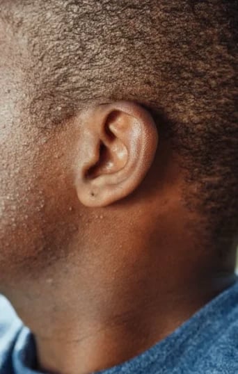
Two consequences matter. First, because there is no cartilage, nothing resists a downward pull. A heavy earring applies constant tension to a small ring of tissue, and soft tissue under sustained tension does what soft tissue always does: it creeps. Over years, the hole migrates downward toward the free edge. Second, because the lobe is a free margin rather than a tethered surface, any scar that crosses that margin at a right angle sits perpendicular to the direction of wound contracture. Scars contract along their length as they mature. A straight scar running from the piercing site down through the rim will shorten as it matures, and it pulls the rim inward with it, producing the small V-shaped notch that is the classic sign of an earlobe closed without regard for geometry.
Those two facts, no cartilage and a free margin, account for most of what goes wrong and most of what a properly planned repair is designed to prevent.
Presentation one: the fully split lobe
This is the one people picture. The piercing tract has torn all the way through the inferior margin, leaving two separate flaps of tissue where there used to be a continuous lobe. It happens in a moment: a hoop caught on a jumper being pulled off, a toddler grabbing an earring, a seatbelt, a hairbrush, a dog. Sometimes it happens slowly, with the tract thinning over months until a minor snag completes what gravity started.
By the time it presents for repair, the two edges are not raw. They have epithelialized. The body has resurfaced each cut edge with skin, which is the key technical point. Two skin-covered surfaces will not knit together no matter how neatly they are held. The epithelialized edge has to be excised on both sides to create fresh, bleeding, raw tissue before anything is closed. This is the single most important step in the whole procedure, and it is the step amateur attempts skip.
Once the tract is freshened, the question becomes what geometry to close it with.
Straight-line closure is the simplest. The freshened edges are approximated in layers and closed directly. It is quick and it works, with one caveat: a straight closure that meets the free rim of the lobe at a right angle leaves a scar oriented exactly along the axis of contracture. As that scar matures over the following months it shortens, and it draws the rim up into a notch. Not everyone notches. Thin lobes, tense closures and patients who scar actively are at higher risk. But it is the predictable failure of the simplest technique, and it is why a straight-line closure at the rim is often modified rather than used in its pure form.
Z-plasty solves the problem by refusing to let the scar run in one direction. Two triangular flaps are raised on either side of the line of closure and transposed, which breaks the straight scar into a zigzag. The functional effect is to lengthen the tissue along the original axis and redirect the line of tension, so contracture no longer pulls in a single direction at the rim. At the free margin, a small Z placed right at the edge is an effective way of preventing notching, because the interdigitating limbs resist the inward pull. The trade-off is a slightly longer total scar and a marginally more complex closure, which in a lobe of this size means a few extra minutes.
L-plasty takes a different approach. Rather than transposing flaps, the closure is carried around the rim: the repair line runs down the lobe and then turns to follow the edge, so that no incision crosses the margin perpendicularly at all. The result is a scar that is partly hidden along the natural contour of the rim and that has no perpendicular limb to contract inward. It is a clean solution for a lobe where the edges are healthy and where tissue can be spared, and it tends to give a very natural rim contour.
Choosing between them is a judgment made at the chair, on that lobe, on that day. It depends on how much tissue has been lost, how thick or thin the lobe is, whether the tear is central or eccentric, and whether the patient scars badly. It is a small decision with a large effect on how the lobe looks in a year, and it reflects training in facial surgery specifically rather than general technique applied to the ear. That background is set out in Dr Doshi's surgical training and credentials, and the meaning of double board certification in facial plastic surgery is worth understanding before choosing anyone to operate on a structure this small.

Presentation two: the elongated or slotted lobe
This one presents differently and is very often misdescribed by the patient as "my earring hole got big."
The hole has not torn through. The inferior margin is intact. But decades of heavy earrings, or a single heavy pair worn daily for years, have stretched the tract downward into an elongated slot. What was a round 1mm opening is now an oval, or a slit, sitting far lower on the lobe than it originally did, sometimes only a millimeter or two of tissue away from the free edge. Studs sit crooked. Hoops rotate. Nothing stays where it is put, because the tissue holding the post no longer grips.
This is a tissue-quality problem as much as a hole problem. The skin around an elongated tract has thinned. The lobe itself is often laxer and less full than it was, partly from the earrings and partly from age, which thins the lobe independently of anything ever having been worn in it.
The repair is different because the objective is different. In a split lobe you are rejoining two separated parts. Here nothing is separated; you are removing an unwanted slot and restoring a competent, appropriately positioned opening or, more commonly, closing it entirely so that a fresh piercing can be placed later.
The usual solution is a wedge excision. The elongated tract, together with the thinned tissue around it, is excised as a wedge, and the resulting defect is closed in layers. Because the wedge removes tissue rather than simply approximating existing edges, the lobe becomes slightly smaller and, importantly, tighter and better supported. Where the elongation sits close to the rim, the wedge is designed so that the closure at the margin is either broken with a small Z or carried along the edge, for exactly the reasons described above.
An additional consideration in this group: because the underlying issue includes tissue laxity, the surgical result can be helped by addressing volume separately. A thin, deflated lobe that has been stretched by jewellery does not regain its former plumpness just because the slot has been closed. That is where non-surgical volume restoration has a genuine, limited role, discussed further below.
Presentation three: the gauged or deliberately stretched lobe
Gauged lobes are the technically most demanding of the three, and the most commonly underestimated.
Stretching is a deliberate, gradual process. Over months or years, a piercing is dilated with progressively larger plugs or tunnels. What is created is not a wound. It is a stable, mature, fully epithelialized tunnel: a cylinder of skin running through the lobe, with a thickened, keratinised lining that behaves like skin because it is skin. Around larger gauges the surrounding tissue also thins and fibroses, so the remaining rim of lobe below and around the tunnel can be a narrow band.
This is why the intuitive approach to gauged ear repair fails. Bringing the two sides of the tunnel together and stitching skin over the top leaves the epithelial lining trapped inside the closure. A buried tract of skin does not disappear. It continues doing what skin does: producing keratin and sebum with nowhere to drain. The result is a firm nodule, sometimes an inclusion cyst, sometimes a persistent draining sinus, sometimes a closure that simply comes apart weeks later because two skin surfaces were never going to fuse. Attempts at home closure, or closure by someone unfamiliar with the anatomy, fail for this exact reason.
The correct operation begins with complete excision of the epithelialized tract. The entire cylinder of skin, front to back, is removed as a unit, along with the thinned and fibrotic tissue immediately around it. What remains is a defect in fresh, healthy, bleeding tissue, which is the only state in which reliable healing can occur.
Reconstruction then becomes a question of what is left. A moderately stretched lobe usually has enough residual tissue for direct layered closure, with the rim geometry planned to avoid notching. A heavily stretched lobe may not: the inferior band may be too narrow to survive as a rim, in which case it is excised and the lobe is reconstructed to a smaller but properly contoured shape. Some large-gauge repairs are best done as a planned two-stage procedure, closing the bulk of the defect first and refining the rim contour once the tissue has settled and softened, typically several months later. Attempting to achieve a perfect rim in one sitting on a lobe with very little spare tissue is the more likely route to a disappointing shape.
Patients with gauged lobes often ask whether the lobe will look like it was never stretched. The honest answer is that it will look like a normal earlobe, usually smaller than the original, with a fine line that fades over months. It will not look untouched under close inspection in raking light. It will look entirely unremarkable in any normal social situation, which is what almost everyone wants. Concerns extending beyond the lobe to the shape or projection of the ear are a separate operation, covered under ear reshaping surgery.
What the procedure is actually like
This is the part that surprises people most, so it is worth being specific.
Earlobe repair is done in the office. It uses local anesthetic only. No general anesthetic, no sedation, no fasting, no anesthetist, no drive home arranged in advance, no operating theatre. One lobe takes roughly twenty to thirty minutes of operating time. Both lobes together generally sit under the hour, allowing for marking and setup.
The lobe is marked with the patient sitting upright, because a lobe hanging in its natural position looks quite different from one lying flat. Local anesthetic is infiltrated, usually lidocaine with epinephrine, which takes effect within a couple of minutes and reduces bleeding in a well-vascularised structure. The only discomfort in the entire procedure is the sting of that injection, lasting a few seconds. After that the lobe is numb and the patient typically talks through the whole thing.
The epithelialized tract or edges are excised. The chosen closure geometry is marked and cut. Deeper tissue is approximated with a small number of buried absorbable sutures, commonly a 5-0 or 6-0 absorbable material, which takes the tension off the skin so the surface closure is sitting on a supported bed rather than holding the lobe together on its own. This layered approach is a large part of why a professionally repaired lobe leaves a fine line rather than a spread scar.
Skin is then closed on both the front and back surfaces with fine non-absorbable sutures, typically 6-0, which are removed at five to seven days. Leaving fine sutures in the face or ear longer than that risks track marks, and the lobe does not need them longer because the buried layer is doing the work.
Because the procedure is this contained, it can very often be performed the same day as the consultation, provided the examination confirms the lobe is straightforward, there is no active infection or inflammation in the tract, and the patient has had the technique and healing timeline explained properly rather than hurriedly. Complex gauged repairs, heavily scarred lobes, revision cases and anyone with a keloid history are better planned as a separate visit. If you want to establish whether your case is a candidate for same-day treatment before traveling in, a virtual consultation will usually settle it from photographs.
The first two weeks, in order
Day of procedure. The local anesthetic wears off over two to four hours. Discomfort is mild and almost always managed with paracetamol or ibuprofen. Normal activity resumes immediately, and people go back to work the same afternoon.
Days one to three. Mild swelling and a pink line. Some bruising is possible on the lobe and occasionally along the jaw below it, because blood tracks downward. The lobe may feel firm and slightly tender. Antibiotic ointment is applied to the suture line twice daily to keep it from crusting.
Days four to seven. Swelling settles noticeably. The line looks sharper and more defined, which is normal. Hair washing is fine from day one or two; the concern is soaking, not water contact.
Days five to seven. Sutures come out. This takes under five minutes and is not uncomfortable. The line at this stage looks thin and pink.
Weeks two to four. The pink line begins to firm up and may look slightly raised. This is the beginning of normal scar maturation and it is the point at which patients most often worry, because the scar looks more prominent at three weeks than it did at one week. That is expected and it reverses.
Months two to four. The peak of the firm, pink phase. Silicone gel or sheeting is worth using from around week three onward, with gentle scar massage once the wound is fully sealed.
Months six to twelve. Softening and fading. Most earlobe scars are pale, flat and difficult to find by twelve months. Full maturation of any facial scar is a year-long process and the lobe is no exception. For scars that remain visible beyond that window, resurfacing with fractional CO2 laser treatment can refine texture and color, and genuinely poor scars from previous non-surgical or badly planned repairs are addressed with scar revision surgery. The broader principles are set out in this guide to scar revision on Long Island.
Re-piercing: the part that decides whether you do this twice
Everything above is the easy part. Re-piercing is where repaired lobes are lost.
The timeline. A repaired lobe should not be pierced again for a minimum of six to eight weeks, and there is a strong argument for waiting three months. The reason is not that the wound is open; it is sealed within two weeks. The reason is tensile strength. A healing wound has recovered only a fraction of its original strength at six weeks, and it continues gaining strength for months as collagen remodels. Putting a needle and then a post, and then the daily tug of an earring, through tissue that is still structurally immature is asking it to fail. Three months is the conservative figure and it is the one worth following if the lobe was thin, the repair was complex, or the tear was a recurrence.
Placement is the critical variable. The single most common reason a repaired earlobe needs a second operation is that it was re-pierced straight through the healed scar line. This makes intuitive sense to almost everyone and it is completely wrong. Scar tissue is not normal tissue. It is less elastic, less vascular, less forgiving under tension, and the piercing tract through it does not mature into a stable, well-supported channel the way a tract through healthy tissue does. A hole placed through the middle of a repair line is placed in the weakest available tissue, in the exact orientation that reopens the original injury. It tears again.
The new piercing must be positioned in healthy tissue away from the repair, usually a few millimeters to one side of the scar and higher on the lobe than the original hole, well clear of the free margin. Higher matters as much as lateral: the original hole was often too low to begin with, which is part of why it failed.
Who should do it. The re-piercing should be performed or directed by the surgeon who did the repair, or by a piercer working to their marked position. The surgeon knows where the scar runs beneath the surface, which is not always obvious from outside once the line has faded. A well-meaning studio piercer aiming for symmetry with the other side will place the hole where the old one was, which is precisely where it should not go.
What to wear afterwards. Small, light studs for the first several months. No hoops, nothing heavy, nothing that a jumper or a child can catch. The original injury was mechanical and the risk factors have not changed. Many patients repair a lobe and then return to the same earrings that tore it.
Keloids and who is at risk
Keloid scarring is the complication that genuinely changes the plan, and the ear is one of the sites in the body where keloids occur most readily. The earlobe in particular is a classic location, often arising months after a piercing rather than after surgery.
A keloid differs from a hypertrophic scar in that it grows beyond the boundaries of the original wound and does not regress. Risk is higher in people with darker skin tones, in those with a family history of keloids, and in anyone who has already formed a keloid anywhere, including on the opposite ear. A previous earlobe keloid is the strongest single predictor of another.
This does not rule out repair. It changes the protocol. A patient with a keloid history is a candidate for planned adjunctive treatment: intralesional corticosteroid injections timed through the healing phase, meticulous tension-free closure with the buried layer taking all the load, silicone therapy started early and continued longer, and close follow-up through the months when a keloid would declare itself. It also changes the re-piercing conversation substantially, and for some patients the right answer is to close the lobe and leave it closed. That conversation is worth having honestly at consultation rather than discovering the issue afterwards.
Volume, aging and the limits of filler
Separately from any piercing history, earlobes age. They thin and lose fat, the skin becomes lax, and the lobe elongates and looks crepey. This happens to people who have never worn an earring, and it is the same volumetric process that affects the rest of the face.
A small quantity of hyaluronic acid filler placed into a thin lobe restores fullness, smooths crepey skin and, usefully, gives a stud something to sit against so it stops drooping forward. It is a five-minute treatment with no downtime, and for the right lobe it is genuinely effective. The dermal filler options suitable for this are the softer products, not structural ones.
The limits need stating plainly. Filler is volume. It is not structure and it is not closure. It will not fix a torn lobe, will not narrow an elongated slot in any durable way, and will do nothing whatsoever for a gauged lobe except add bulk around a tunnel that still needs excising. It is temporary, typically lasting somewhere between six and twelve months in this location. And injecting filler into a lobe that needs surgery simply delays the surgery while adding tissue that may need to be accounted for later. The correct sequence, when both are indicated, is repair first and volume once the scar has matured. Small-scale facial work such as dimple creation surgery succeeds or fails on millimeters, and so does a lobe.
Where earlobe work sits alongside other ear procedures
Patients who dislike the shape of their lobe often dislike the position of the ear as a whole, and the two are worth assessing together even if treated separately. Setback surgery for prominent ears works on cartilage rather than soft tissue and has a different recovery, covered in this breakdown of otoplasty recovery and cost on Long Island. Results across ear procedures sit in the ear surgery gallery.
The practical summary
A torn lobe, a stretched lobe and a gauged lobe are three different problems with three different operations behind them. All three are solved in the office, under local anesthetic, in under an hour, with sutures out in under a week and a scar that is difficult to find within a year. The technical work sits in the geometry at the rim and, for gauged lobes, in removing the epithelialized tract rather than closing over it. Durability is decided afterwards, by waiting long enough to re-pierce and by putting the new hole somewhere other than through the scar.